What is BriteVu
BriteVu is a radiodense contrast agent for use in terminal research subjects and cadavers.
What can it be used for?
BriteVu is primarily used as an intravascular agent to cast the cardiovascular system down to the capillary level. However, BriteVu can be injected into any cavity including the respiratory and gastrointestinal systems, cerebrospinal fluid, body compartments and more. Any animal can conceivably be perfused with BriteVu. BriteVu can also be used to fill compartments of mechanical systems and other non-biologic subjects.
Specific areas where BriteVu can be used in research include the following (and more):
- Anatomic discovery (different species and anatomic locations not previously defined)
- Angiogenesis research
- Artificial organ and biomaterials development
- Cancer development and growth
- Cardiovascular health and disease
- Comparative anatomy
- Delineating vascular anatomy (general)
- Dental health and disease
- Diabetes research
- Embryology/tissue development research
- Geology (pressure perfusion, cavity filling, intercrystalline space, etc)
- Kidney health and disease
- Liver health and disease
- Mechanical and nonbiologic system evaluation (leaks, damage, flow, etc)
- Neurologic health and disease
- Pharmaceutical development
- Organ and system physiology (dermatologic, endocrine, reproductive, etc)
- Organ dysfunction/disease state research (dermatologic, endocrine, reproductive, etc)
- Paleontology research (vascular modeling, cavity perfusions, etc)
- Respiratory health and disease
- Skeletal health and disease
- Space research (effects of space travel on vascular systems and tissues)
- Surgical (vascular and cavity identification)
- Toxicology research
- Transplant research
- Trauma, other tissue injury and healing
How to use it?
PDF: BriteVu Perfusion protocol
*To request a perfusion video demonstration please use the contact form below:
BriteVu advantages
Comparison of Commercially Available Terminal Vascular Contrast Agents
Usage examples:
- Cardiovascular
- Embryology
- Gastrointestinal
- Hepatic
- Infectious Disease
- Neurovascular
- Renal
- Reproductive
- Respiratory
- Special Senses
Cardiovascular: Back
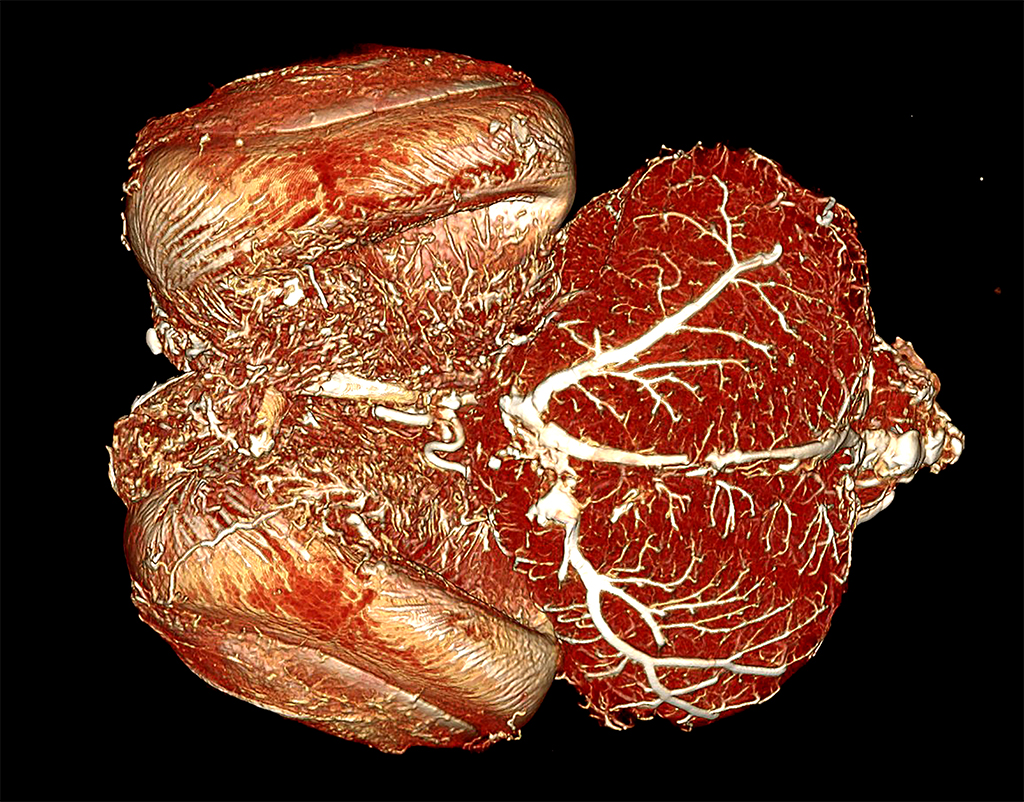
Rat Heart Perfused with BriteVu CT Contrast Agent
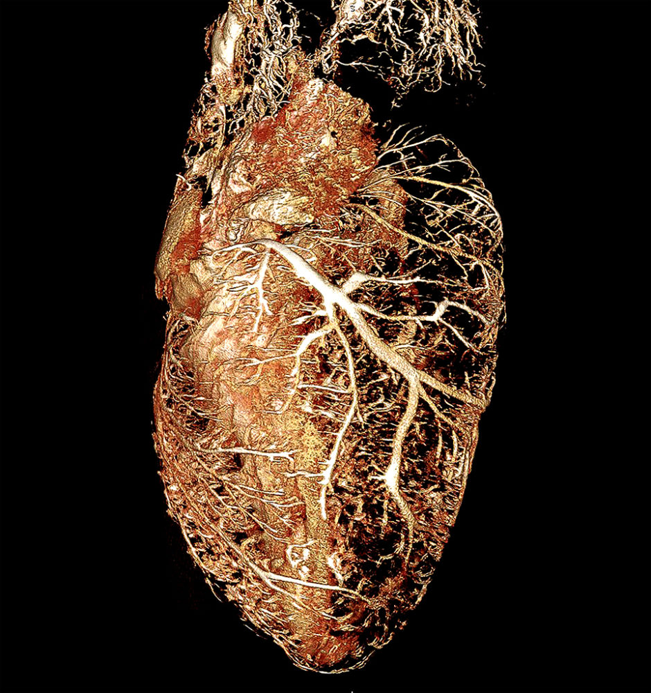
Rat heart perfused with BriteVu contrast agent.
As part of a study at the Head Injury and Vessel Biomechanics Lab at the University of Utah, the whole rat was perfused and individual organs were CT scanned for detailed analysis. The 10 µm scan shows individual blood vessels in the heart muscle and on the outside (especially along the coronary groove). By understanding vascular flow, researchers can determine areas of increased and decreased blood supply to different parts of the heart that may be associated with a large variety of diseases. There is a small amount of motion artifact present.
Alligator Neck and Heart High Radiodensity Contrast Perfusion with BriteVu
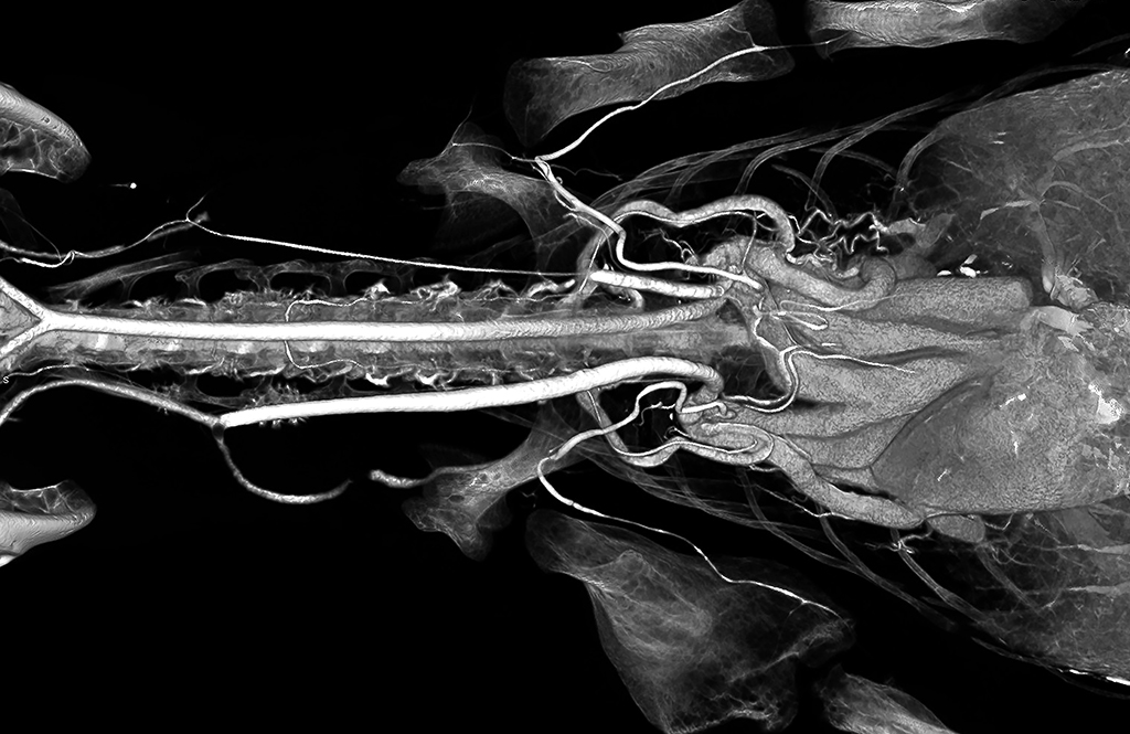
Alligator heart and neck (whole body) BriteVu contrast arteriovenogram.
The alligator was scanned at 100 µm. This is a ventral-dorsal (bottom up) view. BriteVu has proven to be incredibly versatile across multitudes of species. Courtesy of Dr Colleen Farmer, Farmer Lab, University of Utah. Scanned on Siemens Inveon Micro CT.
Embryology: Back
Goose Embryo Perfused with BriteVu While In the Shell
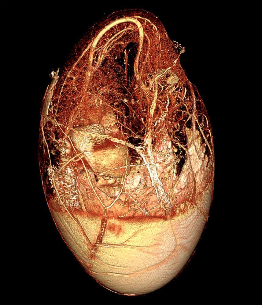
Goose embryo perfused with BriteVu in the egg
Domestic Goose Embryo in Egg (Anser anser domesticus): A goose embryo was perfused while still in the egg. The embryo was an estimated 4 to 7 days from hatch. The portion above the air cell was cut to reveal in the inner shell membranes. Once gently peeled back, the major embryonic vessels were isolated and catheterized. Blood was flushed and replaced with BriteVu contrast agent. A new technique was developed to perform the perfusion. The shell was then partially dissolved to better show the embryo. The egg was scanned using a Siemens Inveon micro CT scanner at 100µm.
Chicken Embryo Perfused with High Radiodensity BriteVu Contrast Media

8 day chicken embryo, vascular perfusion with BriteVu
The embryo was removed from the egg and perfused via cardiocentesis. The developing blood vessels are clearly identified and show how BriteVu can be used in embryology and angiogenesis research. Courtesy of Kirsten Cable & Norman Hu, Department of Pediatrics, University of Utah. 10µm CT scan using Siemens Inveon Micro CT.
Chicken Embryo Perfused with High Radiodensity BriteVu Contrast Media
8 day chicken embryo, vascular perfusion with BriteVu
8 day chicken embryo, vascular perfusion with BriteVu. The embryo was removed from the egg and perfused via cardiocentesis. This image shows a different angle of the same embryo noted above. Courtesy of Kirsten Cable & Norman Hu, Department of Pediatrics, University of Utah. 10µm CT scan using Siemens Inveon Micro CT.
Gastrointestinal: Back
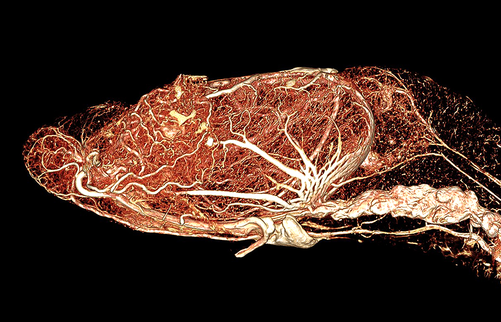
Alligator Distal Colon and Kidney High Radiodensity Contrast Perfusion with BriteVu
An alligator was perfused via the ventral coelomic (abdominal) vein with BriteVu.
Internal organs were removed and CT scanned at 35µm for better visualization. The distal colon and kidney are scanned as one unit. The colon is on the top and terminates to the left and seemingly joins the kidney on the bottom left. Courtesy of Dr Colleen Farmer, Farmer Lab, University of Utah. Scanned on Siemens Inveon Micro CT.
Alligator Stomach and Liver High Radiodensity Contrast Perfusion with BriteVu
An alligator was perfused via the ventral coelomic (abdominal) vein with BriteVu.
Internal organs were removed and CT scanned at 35µm for better visualization. The stomach and liver are scanned as one unit. The stomach is to the right and liver to the left. Courtesy of Dr Colleen Farmer, Farmer Lab, University of Utah. Scanned on Siemens Inveon Micro CT.
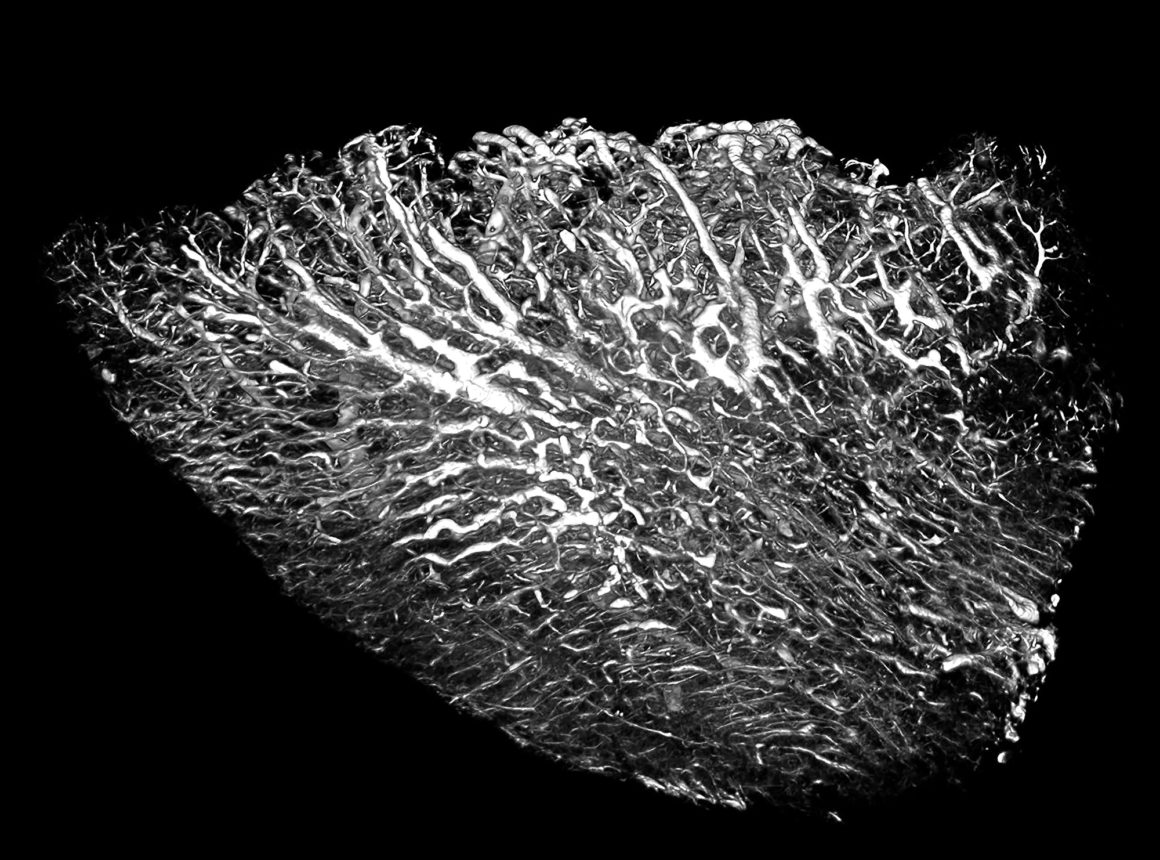
Bearded Dragon Intestines Perfused with High Radiodensity Contrast Media (BriteVu)
Bearded dragon intestines (whole body) perfused with BriteVu contrast.
As seen in this image, BriteVu readily permeates the intestinal vasculature. While not specifically designed for reptiles, BriteVu has shown usefulness in many species in additional to more traditional research animals. The intestines were individually scanned at 35 µm on the Siemens Inveon Micro CT.
Hepatic: Back
Mouse Liver perfused with BriteVu + Metaflow
Mouse liver (whole body) perfusion with BriteVu.
As different researchers have been requesting specific features (when looking at vascular structures), our team has been using a variety of techniques to improve image quality with BriteVu. This image shows the caudal side of a mouse liver perfused with BriteVu and Metaflow (The Dodge Company, http://www.dodgeco.com/). The addition of metaflow improved perfusion, including continuity of filling, of some vascular organs. For more details as to how we used Metaflow with BriteVu perfusions, please ‘Contact’ us. Scanned on Siemens Inveon Micro CT at 35 µm.
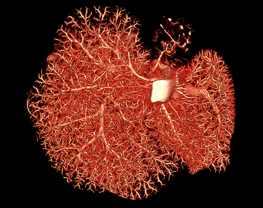
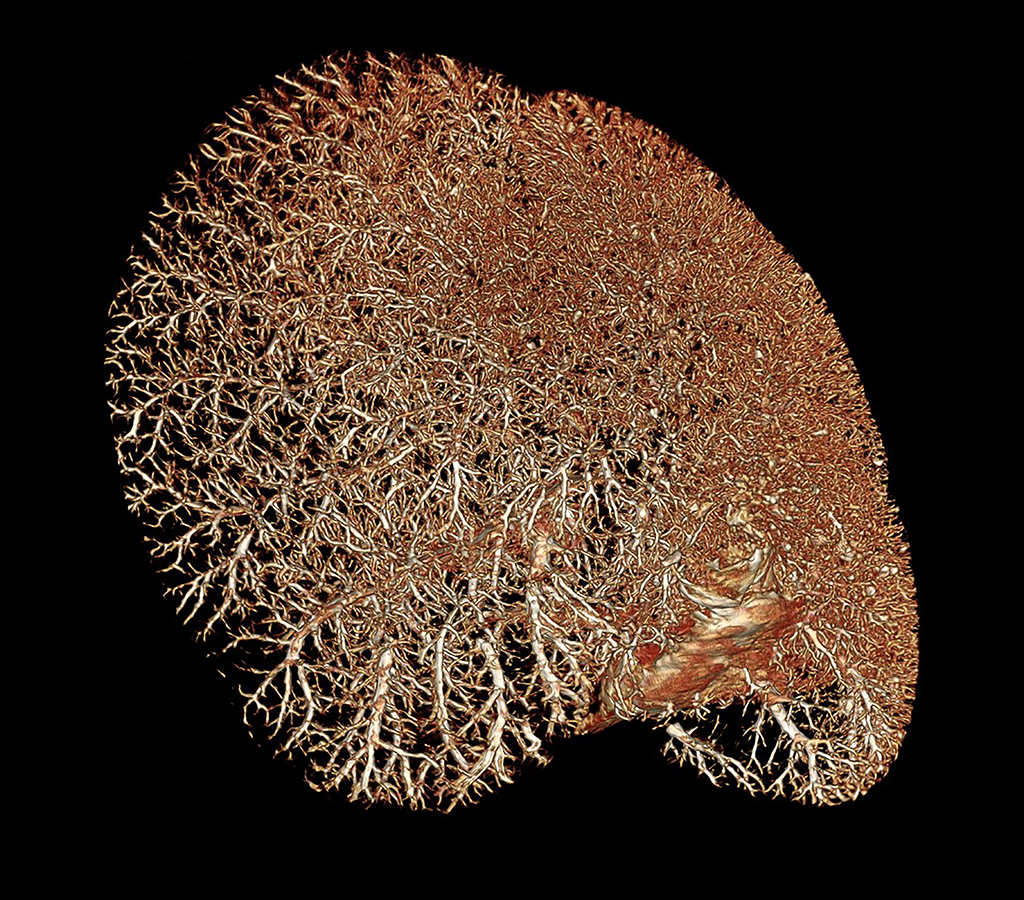
Blue and Gold Macaw Liver High Radiodensity Contrast Perfusion Using BriteVu
Blue and gold macaw liver with BriteVu contrast.
The whole bird was perfused and individual organs scanned. The liver was CT scanned on Siemens Inveon Micro CT at 35 µm slice thickness.
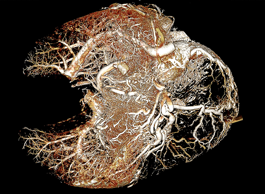
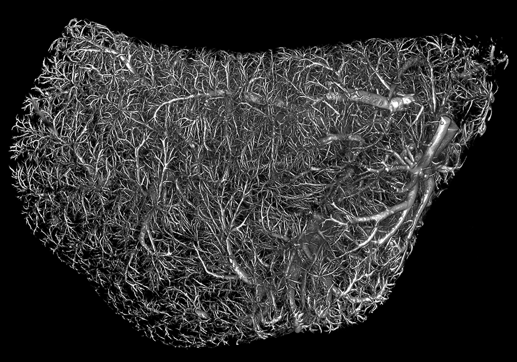
Mouse Liver High Radiodensity Contrast Scan Using BriteVu
Mouse liver (whole body) perfused with BriteVu contrast agent.
The liver was individually CT scanned at 35 µm resolution using Siemens Inveon Micro CT. Because of the highly vascular nature of the liver, the hepatic vasculature is generally very well perfused with BriteVu.
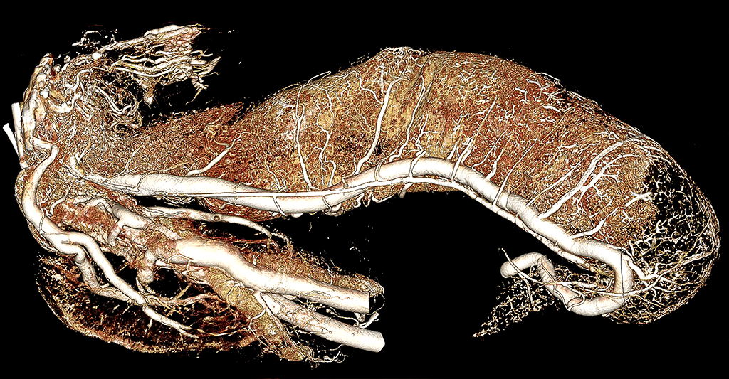
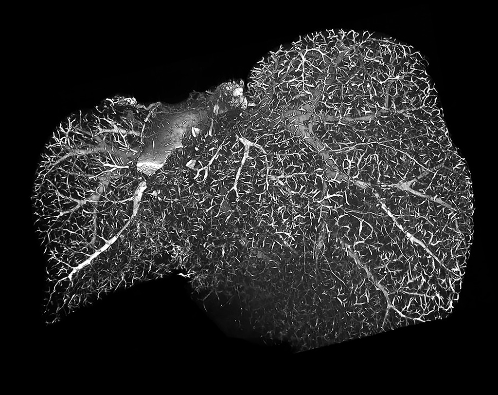
Rat Liver High Radiodensity Contrast CT Angiogram Using BriteVu
Rat liver (whole body) perfused with BriteVu contrast agent.
The liver was individually CT scanned at 70 µm resolution using Siemens Inveon Micro CT. Hepatic studies are common because of the organ’s importance in detoxification, metabolism and much more. BriteVu typically creates beautiful casts of the liver vasculature.
Infectious Disease: Back
Mojave Rattlesnake with Spinal Granulomas and Perfused with BriteVu
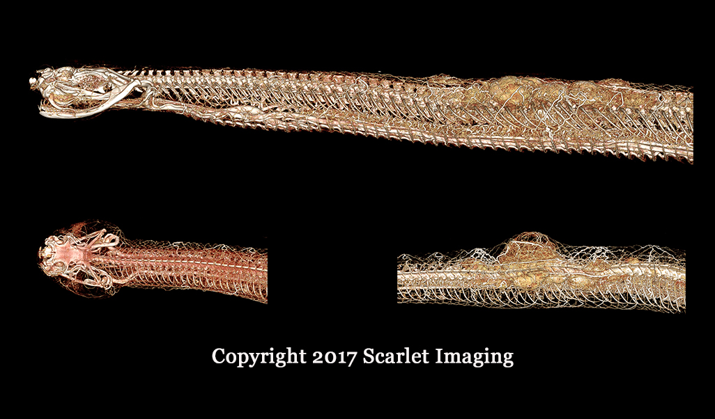
Mojave rattlesnake with BriteVu perfusion
Mojave rattlesnake perfused with BriteVu. The complete vascular perfusion can be seen in the normal head down to the highly abnormal and vascularized infectious granulomas invading the snake’s spinal column. The granulomas are associated with the deformed spinal column. This is a first time look at this type of granuloma vasculature and helps researchers understand how chronic infectious granulomatous diseases get their blood supply. This same information can help lead to better treatments for these types of disorders. Much like cancer, granulomatous diseases (such as tuberculosis) are notoriously difficult to treat. One reason is that it can be challenging to deliver drugs to granulomas. By understanding how blood vessels do, and sometimes don’t, penetrate these lesions gives researchers treatment options. #ScarletImaging #BriteVu #Granulomas
Savannah Monitor Leg Granuloma Perfused with BriteVu CT Contrast Medium
Savannah monitor whole body perfusion with BriteVu CT contrast agent.
This monitor had metabolic bone disease and a large infectious granuloma attached to its entire foreleg (and associated osteomyelitis). Large granulomas in reptiles are poorly vascular making treatment difficult as drugs cannot be easily delivered to the center of the mass. Vascular studies in infectious disease models can be used to help better understand and overcome some of these problems. This is a low resolution scan (0.6 mm CT).
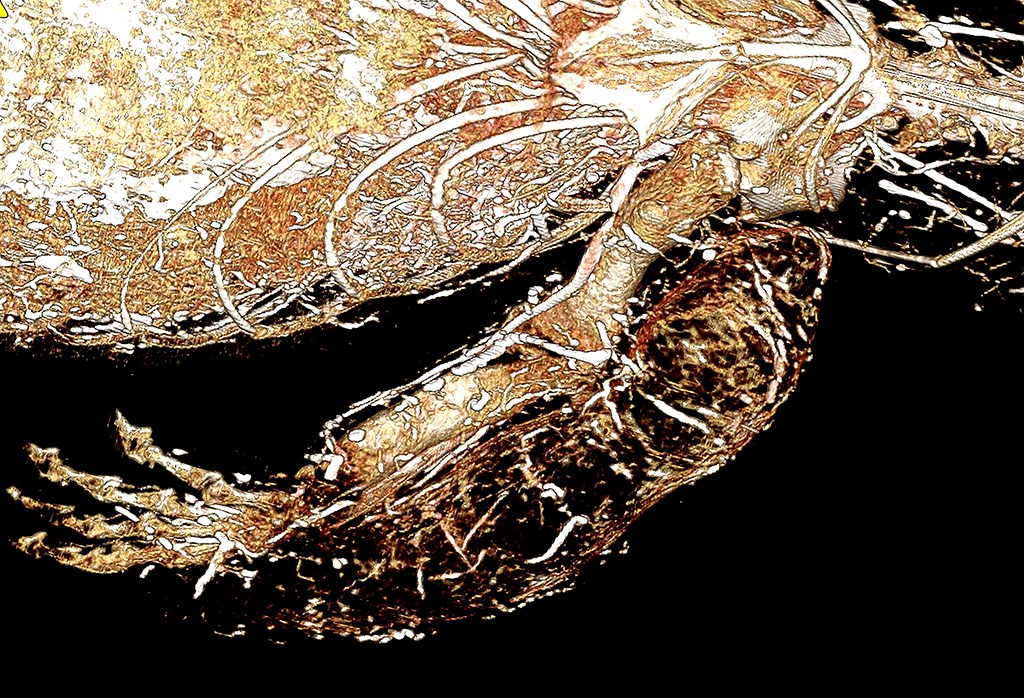
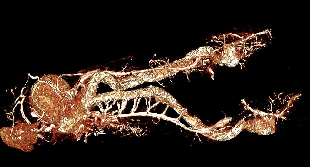
Alligator with Osteomyelitis Perfused with BriteVu (Alligator with osteomyeltitis and BriteVu)
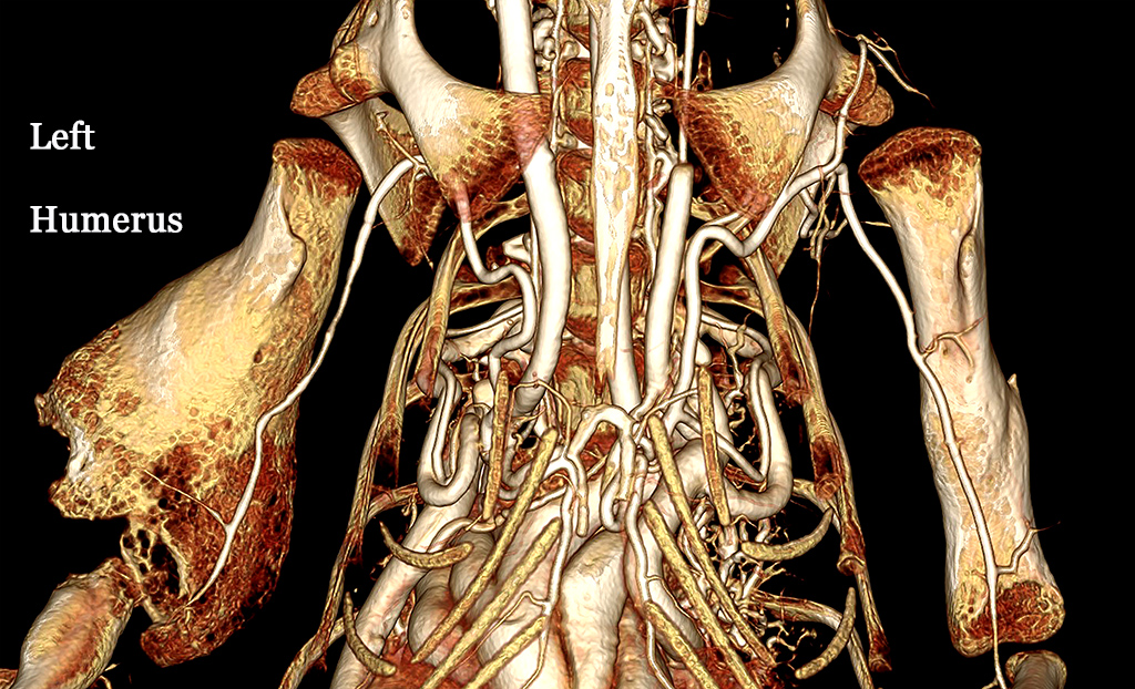
American Alligator whole body perfusion with BriteVu CT contrast agent.
The left humerus has osteomyelitis (bone infection) and has been significantly remodeled (compare with the right humerus). The vasculature to the left humerus has also been altered (decreased) as a result of the chronic infection. This is a ventral-dorsal (or bottom to top) view of the forelegs and heart. Courtesy of Dr Colleen Farmer, Farmer Lab, University of Utah. Scanned on Siemens Inveon Micro CT at 100 µm.
Neurovascular: Back
Pigeon Brain and Eyes BriteVu Contrast Perfusion
Pigeon brain and eyes perfused with BriteVu.
The pigeon was part of an anatomy study evaluating the wing and leg vasculature. The brain and eyes were saved and scanned. This is a dorsal-ventral (or top to bottom) view of the brain (right) and eyes (left). Scanned on Siemens Inveon Micro CT at 35 µm.
BriteVu Perfused Rat Brain
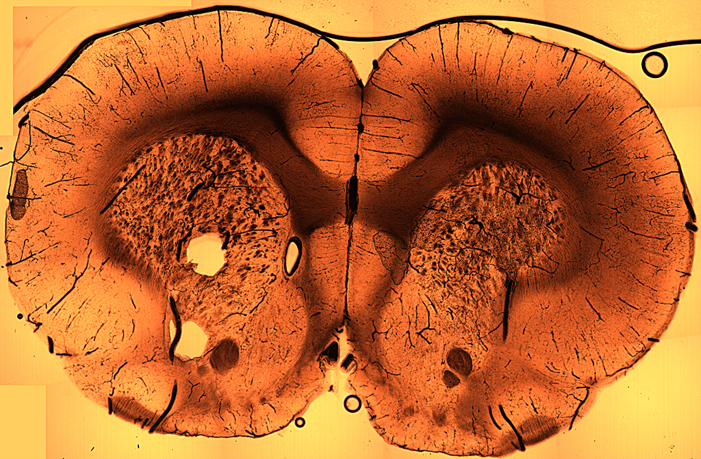
Rat brain frontal plane slice (photo).
Stewart Yeoh from the Head Injury and Vessel Biomechanics Lab at the University of Utah provided this image of a BriteVu perfused rat brain. The dark lines represent blood vessels in the brain. This is a 200 µm slice with no stain, filters or special lighting (basic white light used). From the orientation of the vessels you can get a sense of how blood flows within the brain. Once BriteVu has solidified, tissues can be stored in formalin or other fixatives and processed later for a variety of imaging options.
Human Cadaver Head Perfused with BriteVu
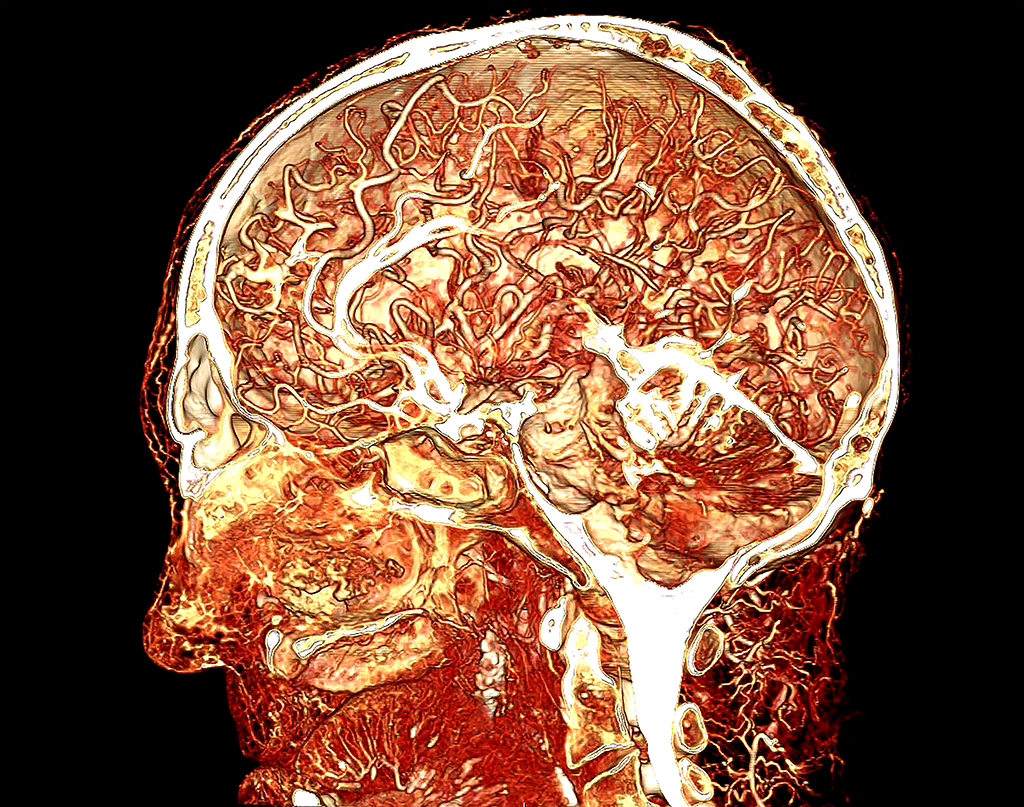
Human cadaver head perfused with BriteVu.
The perfusion was performed by Dr. Bruce Wainman, Education Program in Anatomy, McMaster University. This sagittal section shows the rich blood supply within the human brain. This complexity is difficult to appreciate from a picture alone and is best evaluated using 3D viewing software. While there are numerous reasons to study the neurovasculature in animals, this particular study shows how BriteVu can be used in cadavers. The scan was performed on a GE clinical CT scanner at 1 mm slice thickness.
Renal: Back
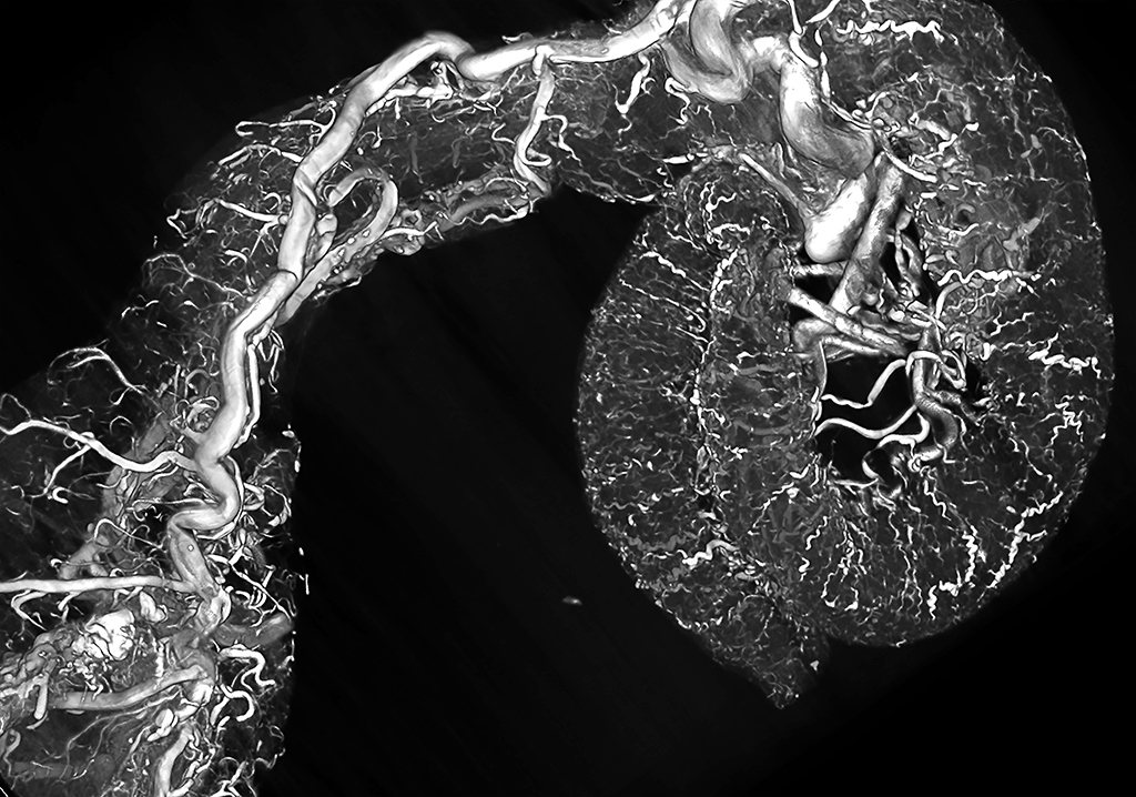
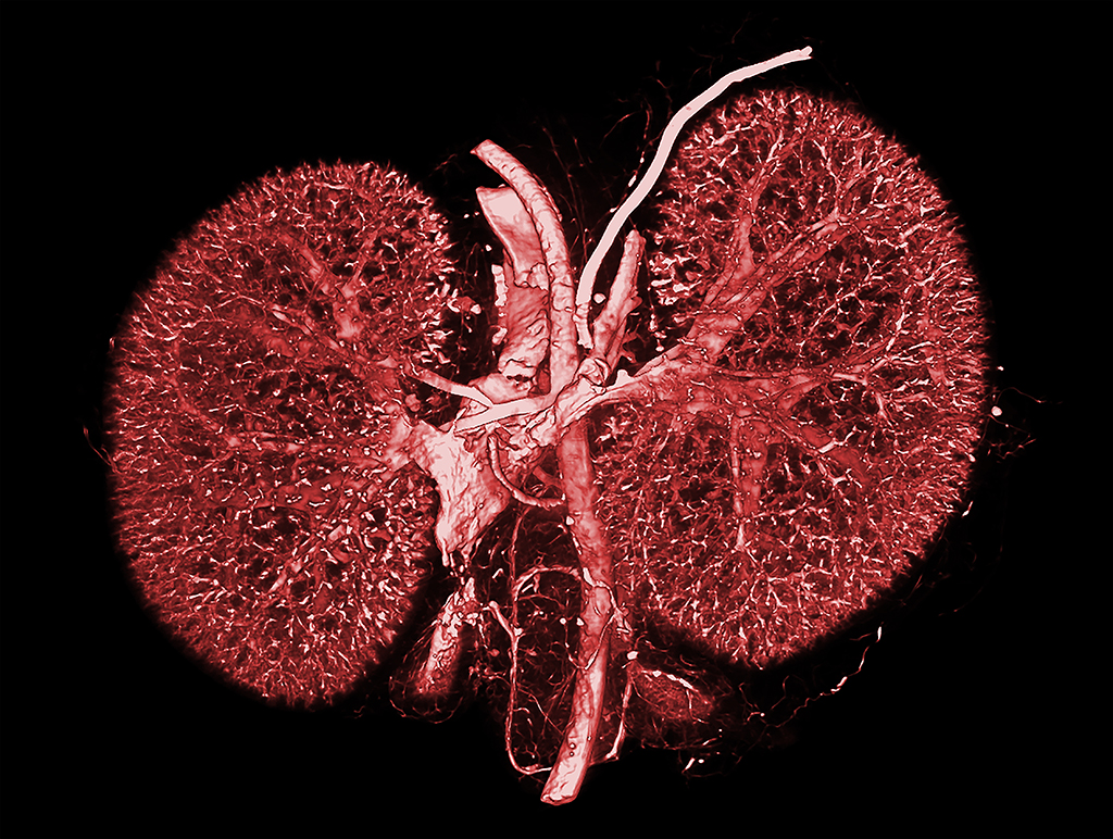
Rat Kidney BriteVu Perfusion

BriteVu perfused rat kidney CT scanned at 30 µm.
Courtesy of the Head Injury and Vessel Biomechanics Lab at the University of Utah. The whole rat was perfused for a brain vasculature study. However, all of the tissues were well infiltrated with BriteVu creating incredible perfusions throughout the body. Scanned on Siemens Inveon Micro CT.
Pigeon Kidneys and Ovary Perfused with BriteVu

Pigeon kidneys and ovary (whole body) perfusion with BriteVu.
This is a ventral view of the kidney. The ovary ‘grape cluster’ can be seen to the right of the image. Major vessels including the common iliac vein and small unnamed intrarenal vessels are readily seen. This information is highly beneficial when studying blood flow, angiogenesis and more through the kidneys (and ovary). Scanned on Siemens Inveon Micro CT at 35 µm.
High Radiodensity Contrast CT of Mouse Kidneys Perfused with BriteVu
Mouse kidneys perfused with BriteVu
The adrenal glands (and their vasculature) are located at the bottom of the image next to the large veins. Scanned on Siemens Inveon Micro CT.
High Radiodensity Contrast CT of Mouse Kidneys Perfused with BriteVu (Invert)
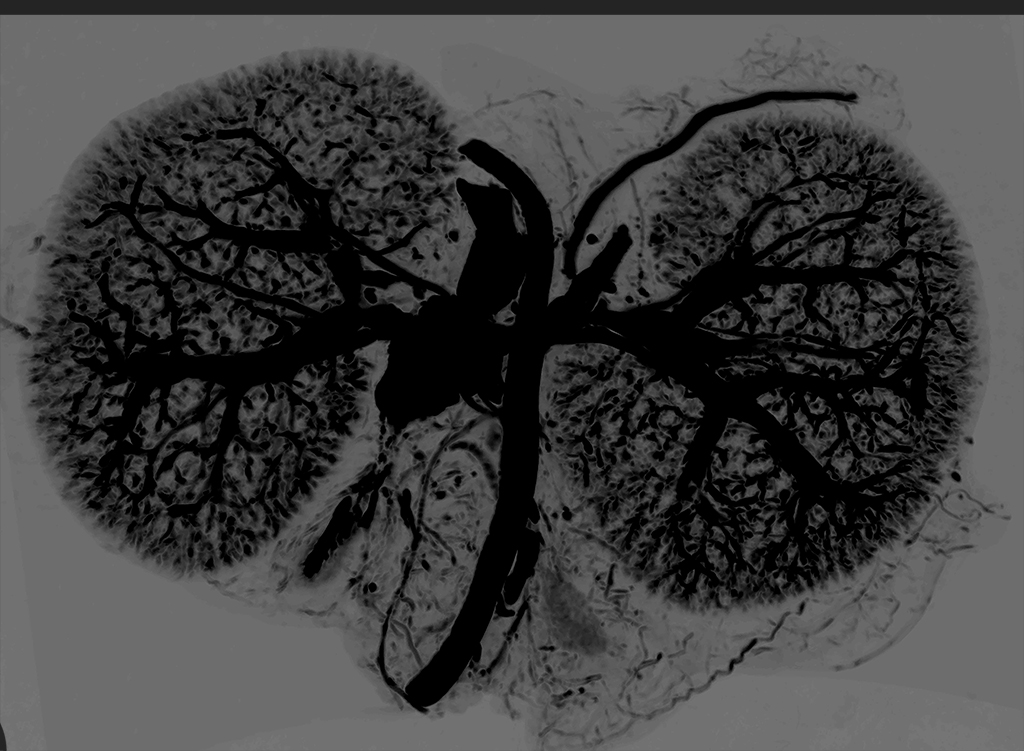
Mouse kidneys perfused with BriteVu (whole body perfusion).
The tissues were CT scanned at 35 µm and the image was then inverted (same image as above). Scanned on Siemens Inveon Micro CT.
Argentine Black and White Tegu Kidneys and Ovary
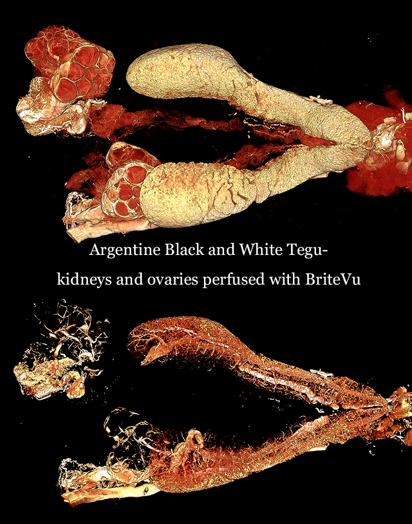
Tegu kidneys and ovaries perfused with BriteVu
The kidneys and ovaries of the Argentine black and white tegu have been perfused with BriteVu and CT scanned at 100 µm. The ovarian follicles are readily visible in the top view. Both images show the dorsal surfaces of the ovaries (left) and kidneys (right). By adjusting the image intensity and contrast, the kidney vessels can be seen below the surface. Scanned on Siemens Inveon Micro CT.
Reproductive: Back
Mouse Ovaries, Uterus and Urinary Bladder Perfused with BriteVu
Mouse uterus, ovaries and urinary bladder perfused with BriteVu
The female mouse reproductive tract and urinary bladder have been perfused with BriteVu. The ovaries are seen to the right and connect to the uterine body and urinary bladder (to the left) via the two uterine horns. The tissues were CT scanned at 25 µm on the Siemens Inveon Micro CT.
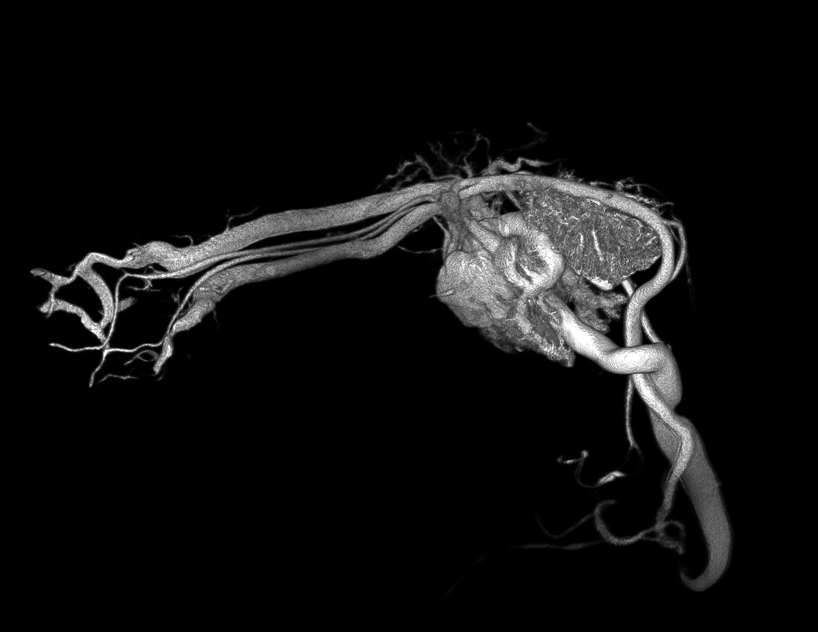
Rat Ovary Perfused with BriteVu

BriteVu Perfused Rat Ovary
Whole rat perfusion with BriteVu. The ovarian blood supply (top left-center) and its closely related uterine vasculature (lower right) are readily seen. The tissues were CT scanned at 35 µm on the Siemens Inveon Micro CT.
Rat Testicle Perfused with BriteVu
Rat testicle perfused with BriteVu.
The epididymis and vascular pampiniform plexus are both readily visible. Courtesy of the Head Injury and Vessel Biomechanics Lab at the University of Utah.
Rat Testicle Perfused with BriteVu (Color Enhanced)
Rat testicle perfused with BriteVu.
This color enhanced image helps demonstrate the significant amount of vasculature present within the testicle. Some species, like birds and reptiles, can dramatically increase the size of their testicles when reproductively active. This tissue hypertrophy (growth) requires recruitment of increased blood supply and can be visualized using BriteVu. Courtesy of the Head Injury and Vessel Biomechanics Lab at the University of Utah.
Respiratory: Back
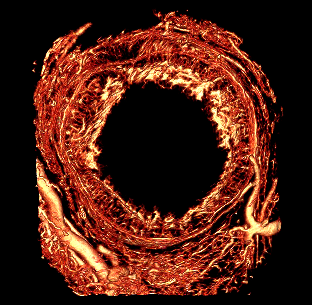
Pigeon Lung High Radiodensity Contrast Perfusion Using BriteVu
Pigeon lungs (whole body) BriteVu arteriovenogram.
The lungs were removed and individually scanned on Siemens Inveon Micro CT at 35 µm.
High Radiodensity Contrast Scan of a Rat Lung Using BriteVu

Rat lung perfused with BriteVu.
Vascular anomalies in the lungs can correlate with numerous diseases, exposure to toxins and environmental conditions, individual variation and more. Courtesy of C. Happé / VU University Medical Center Amsterdam / Department of Pulmonology. CT scan resolution 36 µm. The contrast agent is BriteVu.
Mouse Lungs High Radiodense Contrast Perfusion Using BriteVu
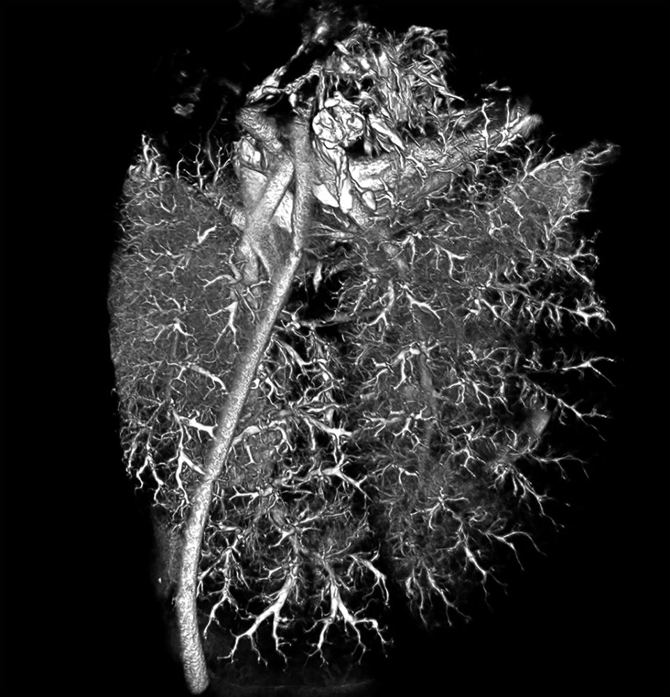
Mouse lungs (whole body) perfused with BriteVu contrast agent.
The lungs are usually the first organs to become perfused via a cardiac puncture creating outstanding images when using BriteVu. Lungs were individually CT scanned at 35 µm resolution on the Siemens Inveon Micro CT.
Rat Lungs High Radiodensity Contrast CT Using BriteVu
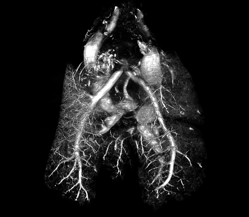
Rat lungs perfused with BriteVu and CT scanned at 70 µm on the Siemens Inveon Micro CT.
The lungs were scanned intact and still connected to each other via the pulmonary arteries coming off the heart. This is a dorsal-ventral view.
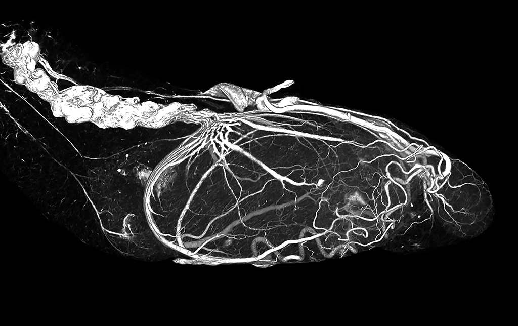
Alligator Lungs Perfused with BriteVu
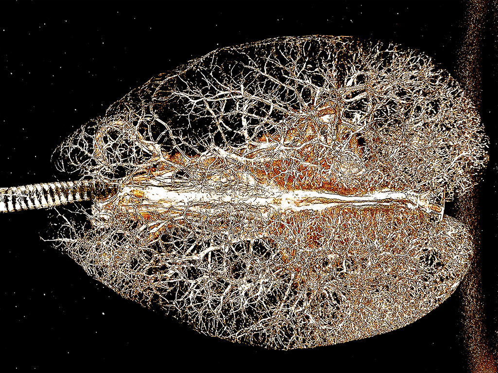
American Alligator heart and lungs perfused with BriteVu.
This is a dorsal-ventral (or top to bottom) view of the lungs. The heart vasculature is partially visible through the lung vessels. The trachea (windpipe) is seen entering the lungs from the left side of the image. Courtesy of Dr Colleen Farmer, Farmer Lab, University of Utah. Scanned on Siemens Inveon Micro CT at 35 µm.
Special Senses: Back
Rat Eye BriteVu Arteriovenogram
Rat (whole body) iris perfused with BriteVu.
Window to the soul. An arteriovenogram reached the eye of a rat and, combined with micro CT, produced a detailed map of the blood supply to and within the eye. The central (black) portion represents the pupil which is immediately outlined by the blood supply to the iris. The back half of the eye has been digitally removed to create a clearer picture. CT scan at 10µm. Courtesy of the Head Injury and Vessel Biomechanics Lab at the University of Utah. Scanned on Siemens Inveon Micro CT
Human Ear BriteVu Perfusion
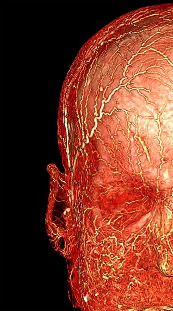
Human cadaver head perfused with BriteVu.
The external ear is isolated showing the rich blood supply to the pinna. The eye socket is visible just to the right of the ear in this picture. This particular study shows how BriteVu can be used in cadavers. The scan was performed on a GE clinical scanner at 1 mm slice thickness.
Mouse Head and Tongue BriteVu Perfusion
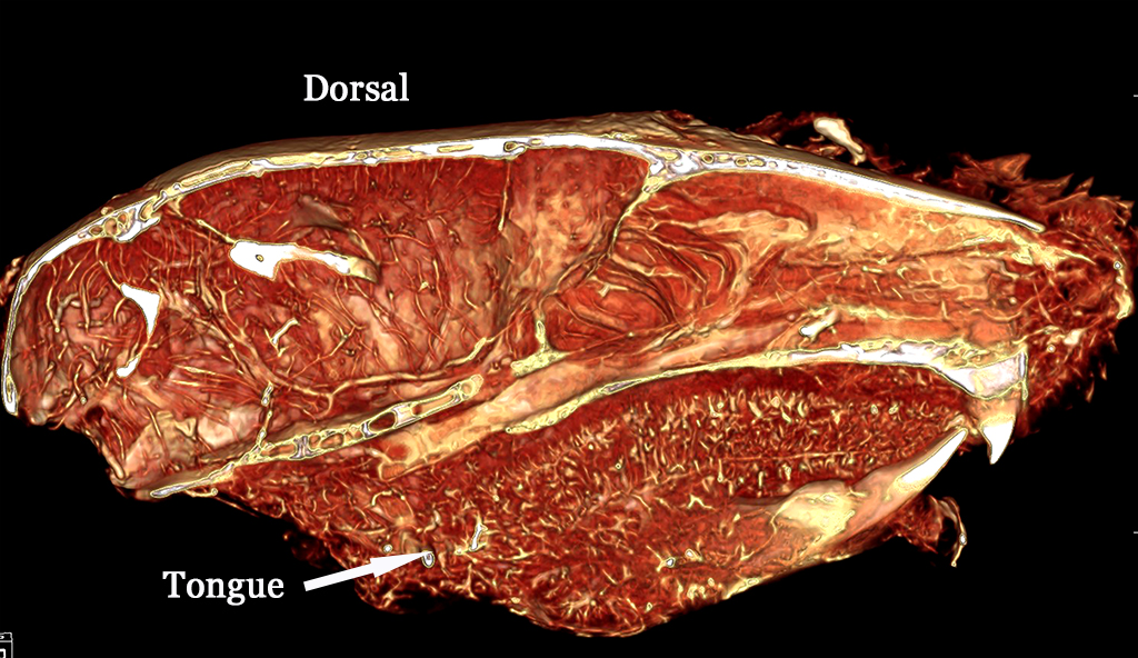
Mouse head perfused with BriteVu and Dawn Ultra.
Dawn can be added to BriteVu solution to improve perfusion in some tissues. In this example, the tongue was extremely well vascularized and can be readily seen on the sagittal view of the head. For more details as to how we used Dawn Ultra with BriteVu perfusions, please Contact us. Scanned on Siemens Inveon Micro CT at 30 µm.